An inner ear infection can cause symptoms and signs for example a severe ear dizziness vertigo nausea and vomiting and vertigo. An inner ear infection or otitis interna is caused by viruses or bacteria and can occur in both adults and children.

Anatomy Of The Inner Ear A Schematic Depiction Of The Inner Ear Which Download Scientific Diagram
THE EAR Outer Ear Middle Ear Inner Ear SlideShare uses cookies to improve functionality and performance and to provide you with relevant advertising.
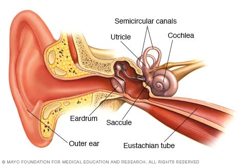
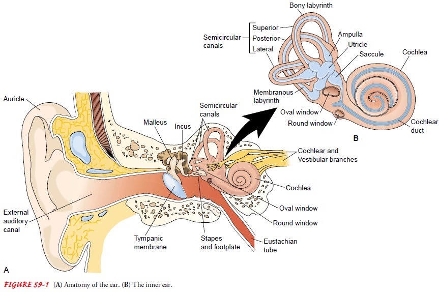
Inner ear anatomy utricle. At first glance the machinery for hearing and balance appears very crude. Anatomy of Ear External ear 1Auricle pinna 2External auditory canal 3Tympanic membrane Ear drum Inner ear 1Semicircular canals 2Vestibule 3Cochlea Middle ear 1Auditory Ossicles 2Oval window 3Eustachian tube 4. The inner ear is a minute complex fluidfilled structure surrounded by a bony labyrinth and located deep in the temporal bone.
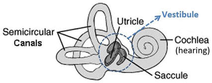
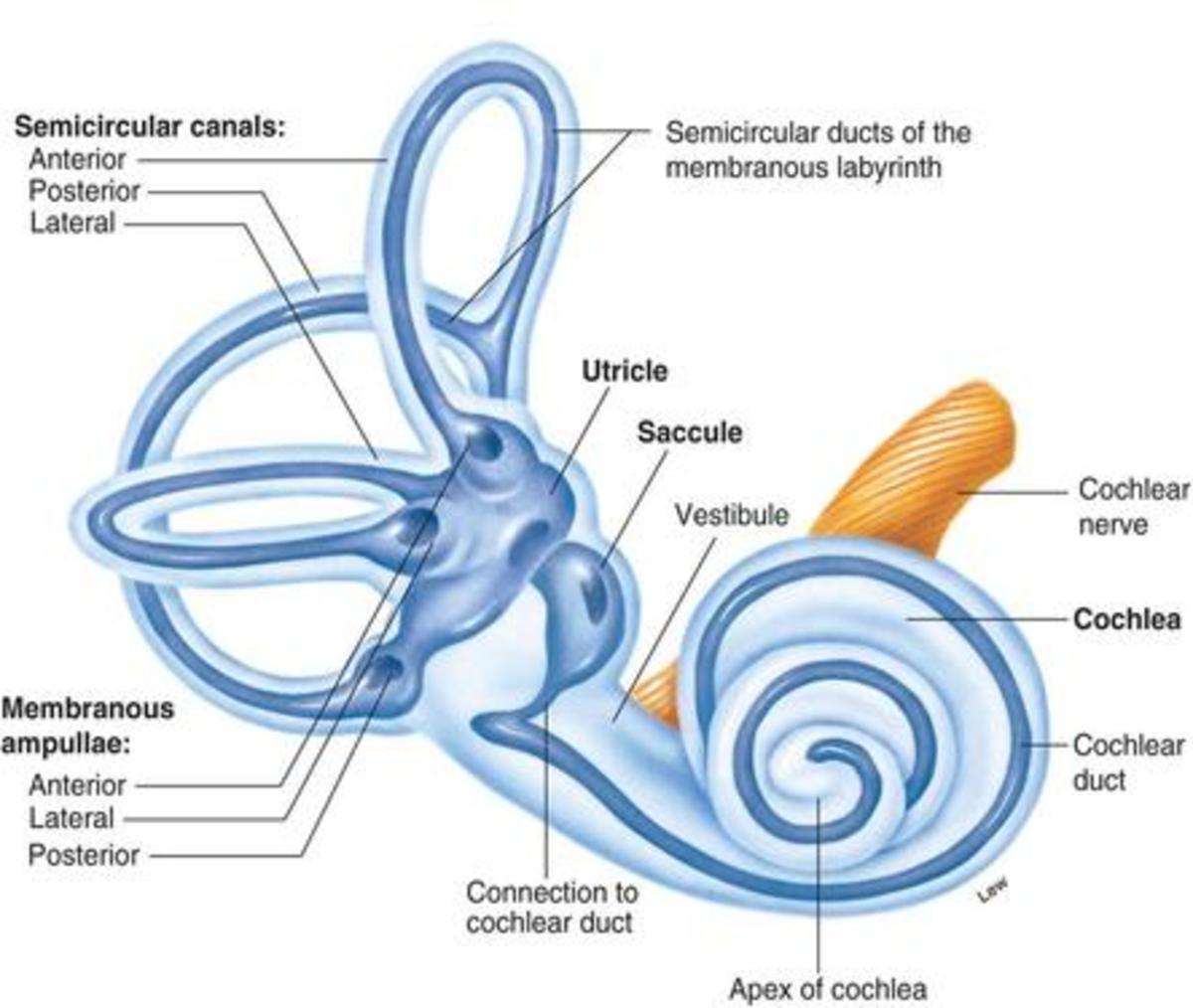
The temporal bone is divided into several main partsportions 1-3. It has an ovular irregular shape and receives drainage from the semicircular ducts. The cochlea corresponds to the acoustic end organ and the vestibular end organs consist of the three semicircular canals with their ampullary tissue the saccule and the utricle.
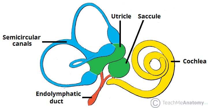
Sense of balance and position is regulated by structures in the inner ear most notably the semicircular canals and the utricle and saccule in the vestibule. Your inner ear is the deepest part of your ear. The ear is the organ that enables hearing and in mammals body balance using the vestibular systemIn mammals the ear is usually described as having three partsthe outer ear the middle ear and the inner earThe outer ear consists of the pinna and the ear canalSince the outer ear is the only visible portion of the ear in most animals the word ear often refers to the external.
In mammals it consists of the bony labyrinth a hollow cavity in the temporal bone of the skull with a system of passages comprising two main functional parts. The middle ear and the internal or inner ear. The vestibular system of the inner ear provides a persons sense of balance and awareness of spatial orientation to coordinate body movements.
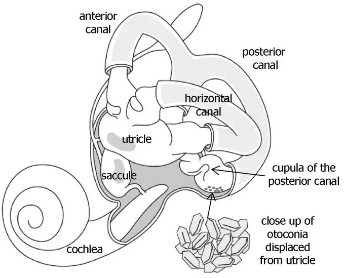
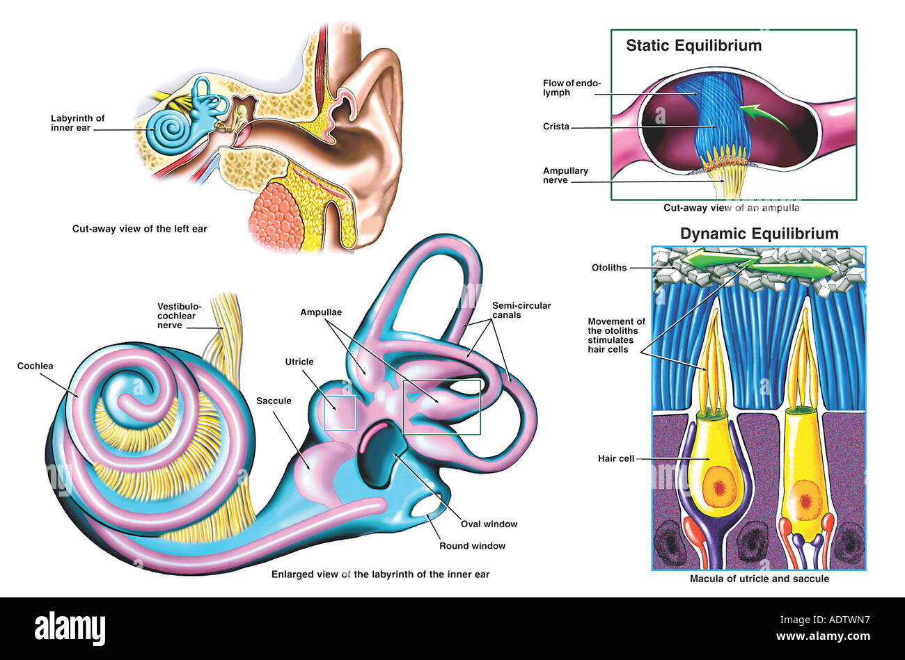
Inner Ear Anatomy. The Canalith Repositioning Procedure is also known as the Epley maneuver BPPV Benign Paroxysmal Positional Vertigo occurs as a result of displaced otoconia which are small crystals of calcium carbonate also referred to as otoliths or canaliths that are normally attached to the otolithic membrane in the utricle of the inner ear. Vestibular nerve enlarges to form the vestibular ganglion which then splits into superior and inferior parts to.
Anatomically the ear is divided into three major areas. Squamous part temporal squama. The inner ear has two special jobs.
The utricle is the larger of the two sacs and is located in the posterior-superior part of the vestibule. The temporal bone is one of the most important calvarial and skull base bones. It changes sound waves to electrical signals nerve impulses.
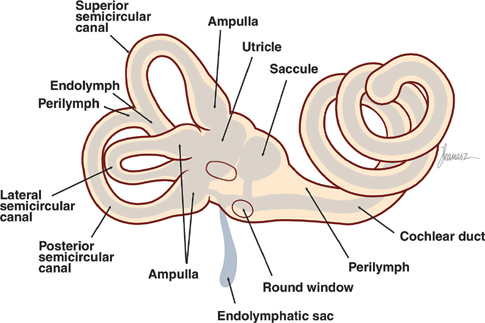
Gelatinous substance in the utricle and saccule of the inner ear that contains calcium carbonate crystals and into which the stereocilia of hair cells are embedded outer segment in the eye the section of a photoreceptor that contains opsin molecules that transduce light stimuli. Petrous part petrous pyramid. The anatomy of the vestibular apparatus balance portion of the inner ear The peripheral vestibular system the non-auditory portion has three semicircular canals that detect angular motion and two otolithic organs the utricle and saccule which detect linear acceleration and deceleration see figure 3.
NOTES NOTES ANATOMY PHYSIOLOGY NERVOUS SYSTEM ANATOMY PHYSIOLOGY osmsitnervous-system-anatomy-physiology THE NERVOUS SYSTEM Network of brain spinal cords nerves Sensoryafferent integrative motorefferent functions Sensoryafferent Receptors monitor external internal environment Conscious stimuli eg. The three semicircular canals correspond to the three dimensions x y and z and connect to the utricle at an ampullaa widening of the canal. An inner ear infection also may cause inflammation of the inner ear or labyrinthitis.
The inner ear is innervated by the vestibulocochlear nerve CN VIIIIt enters the inner ear via the internal acoustic meatus where it divides into the vestibular nerve responsible for balance and the cochlear nerve responsible for hearing. Anatomy of the Ear. The temporal bone is situated on the sides and the base of the cranium and lateral to the temporal lobe of the cerebrum.
If you continue browsing the site you agree to the use of cookies on this website. The inner ear internal ear auris interna is the innermost part of the vertebrate earIn vertebrates the inner ear is mainly responsible for sound detection and balance. The external or outer ear.
The cochlea dedicated to hearing. This allows the brain to hear and understand sounds. The bony labyrinth is formed from the vestibule.
Anatomy of the inner ear. Anatomy of Ear Ear is divided into three main regions External ear Middle ear Inner ear 3.

Utricle Anatomy Britannica
Vestibular System Neupsy Key
Benign Paroxysmal Positional Vertigo Bppv Veda
Inner Ear And Balance Mayo Clinic
How Does The Ear Help To Maintain Balance And Equilibrium Of The Body Owlcation

Inner Ear Illustrations Image Radiopaedia Org

Anatomy Of The Left Inner Ear Showing Displaced Otoconia From The Download Scientific Diagram

Ear Education Tampa Bay Hearing
The Inner Ear Bony Labyrinth Membranous Labryinth Teachmeanatomy
Anatomy Of The Inner Ear Stock Photo Alamy

Utricle Saccule Utricle And Saccule Human Anatomy And Physiology Ear Anatomy Eye Anatomy
Anatomy Of The Inner Ear

Anatomy Of The Inner Ear Enteducationswansea

Utricle And Saccule Anatomy General Pathology And Diseases

Anatomy Of The Otoliths

Inner Ear Wikipedia

Vestibular Information Biology For Majors Ii
Peripheral Vestibular System Avora Health

Anatomy Of Inner Ear It Consists Of Six Mechanoreceptor Structures Download Scientific Diagram

0 comments:
Posting Komentar